Chordoma - Causes, Symptoms, Treatment


Introduction
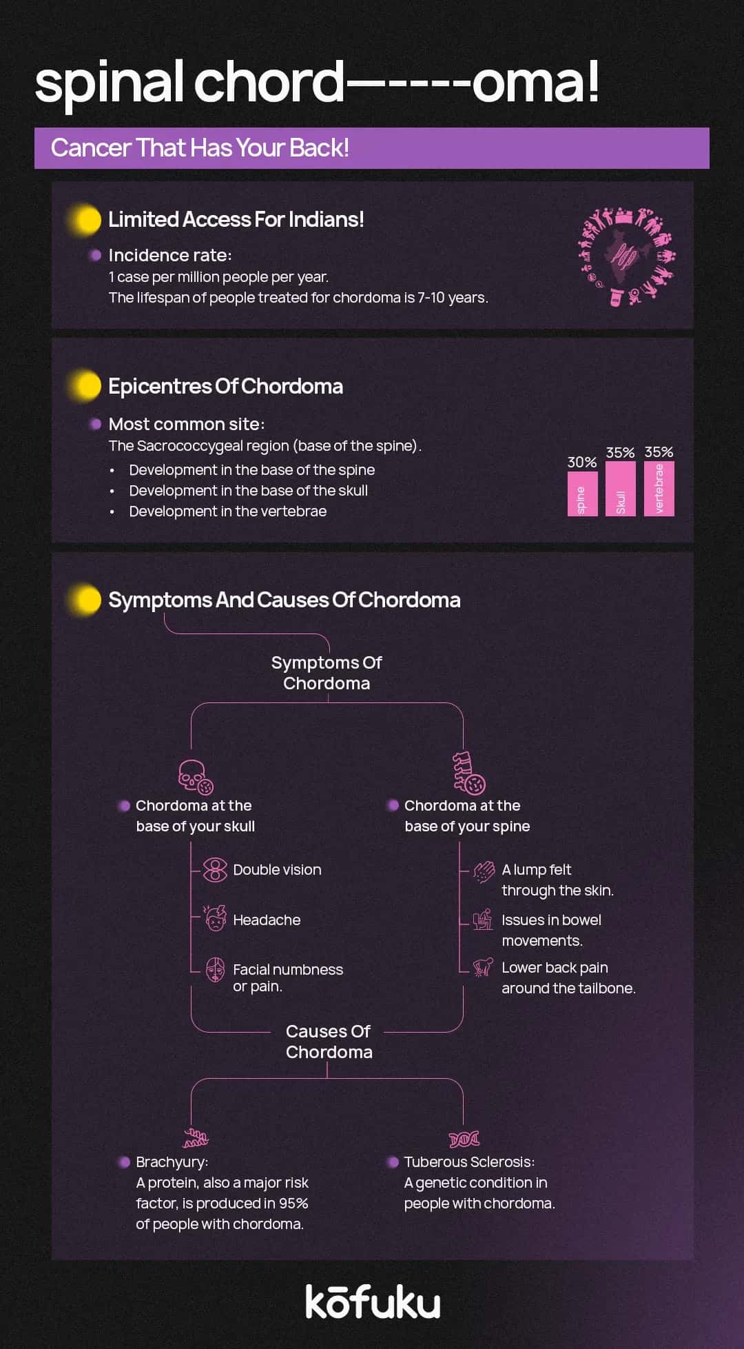
Rare cancers - cancers that don’t have a very high incidence rate, at least in our country. What makes them special is that they do occur, with whatever minimal incidence rate. One such cancer is chordoma.
A slow-growing cancer of tissue found inside the spine, chordoma can occur anywhere along the spine, most often found near the tailbone or where the spine meets the skull. Chordoma is also known as notochordal sarcoma.
Chordomas form the left-over cells that were vital in the development of the spine prior to birth, also called notochord cells. When these cells don’t vanish after birth, they can turn into chordomas.
The rate of growth of chordomas is quite slow. Most people don’t notice any physical changes for years. When symptoms do crop up, it can take a while for the chordoma to be found and diagnosed.
Most people get diagnosed with chordoma in their 50s and 60s. Pediatric chordomas comprise 5% of all chordoma diagnoses. Females get diagnosed a little more than males in childhood. Males are usually diagnosed as adults.
Chordoma is an extremely rare variety of bone cancer that occurs usually in the bones of the skull or the spine. It usually forms where the skull sits on top of the spine or at the bottom of the spine.
Chordoma starts in cells that once made up a collection of cells in the developing embryo which go on to become disks of the spine. Most of these cells disappear by the time of your birth or soon after. However, sometimes, a few of these remain and in the rare occasion - they can become cancerous. Chordoma usually happens to adults aged between 40-60, although it can happen at any age. Chordoma usually grows slowly. It can get tough to treat because it is very close to the spinal cord and other important structures like the arteries, brain or nerves.
Types of Chordoma
Chordoma is differentiated depending on the type of tumour, size, location and pathology.
Conventional chordoma
This is a classic case of tumour which accounts for around 70-80% of the most common chordoma.
Dedifferentiated chordoma
This kind of tumour is aggressive and accounts for less than 5% of all tumours. It is usually found near the base of the spine, but can happen anywhere on the spinal cord or the skull.
Chondroid tumours
These kinds of tumours pop up near the base of the spine, and consist of 10-25% of all tumours. They usually come up and grow because of abnormal cartilage tissues and the abnormal notochord. Its features are just like chondrosarcoma - a variety of bone cancer.
Symptoms of Chordoma
A slow-growing tumour, chordoma doesn’t showcase any symptoms till the patient is of a certain age. Because the tumour is located near the nerves, most of these symptoms are neurological, like.
-
Headaches - Cluster headaches.
-
Pain - arms, legs and back
-
Visual issues - double vision, nerve or muscle weakness.
-
Paralysis of the facial nerve.
-
Fatigue and tiredness.
Because the tumour can grow anywhere near the skull or the spine, the signs and symptoms vary significantly. Such signs and symptoms are quite commonplace and can happen in other conditions as well which makes it quite hard to detect the tumour.
Symptoms of tumour at the base of the spine
-
Lower back pain
-
Inflammation and tenderness.
-
Lump in the back.
-
Constipation and loss of bladder control.
Symptoms of tumours in the neck
-
Pain and inflammation.
-
Breathing obstruction.
-
Dysphagia (difficulty swallowing)

Diagnosis of Chordoma
Removing a sample of cells for testing in the lab (also called a biopsy). A biopsy is a procedure to remove a sample of suspicious cells for laboratory testing. In the lab, well-qualified doctors known as pathologists look at these cells under a microscope to find out whether or not cancer cells are present.
Figuring out how the biopsy needs to be performed requires proper planning by the medical team. Doctors have to do this biopsy in a way that won’t mess with future surgery to get rid of the cancer. For this reason, ask your doctor to refer you to a team of experts who have a lot of experience in treating chordoma.
Get more detailed imaging - Your healthcare provider might want you to take up some imaging tests to assist with visualising chordoma and figuring out whether it has spread beyond the spine or skull base. Tests might include an MRI or a CT scan.
Once you receive a diagnosis of chordoma, your doctor will come up with a treatment plan tailored to your requirements in consultation with experts in ear, nose and throat medicine (otolaryngology), cancer (oncology) and radiation therapy (radiation oncology) or surgery. Your care team might bring in experts in endocrinology, rehabilitation and ophthalmology as required.
Treatment
Chordoma treatment depends on the size and location of the cancer, as well as whether it has invaded nerves or other tissue. Options might include surgery, radiation therapy, targeted therapies and radiosurgery.
Treatment for chordoma in the sacral spine.
If the chordoma impacts the lower portion of the spine (sacrum), treatment options include.
Surgery
The aim of surgery for a sacral spine cancer is to get rid of all of the cancer and some of the healthy tissue which surrounds it. Surgery might be tough to pull off because the cancer is near critical structures, like blood vessels and nerves. When the cancer can’t be removed completely, surgeons might try to get rid of as much as possible.
Radiation therapy
Radiation therapy uses high-energy beams, like X-rays or protons to kill cancer cells. During radiation therapy, you like supine on a table as a machine navigates around you, directing the radiation beams to precise points on the body.
Radiation therapy might be used post surgery to shrink a cancer and make it easier to remove. It could also be used post surgery to polish off any cancer cells that remain. If surgery is not an option, radiation therapy could be the way to go instead.
Treatment with newer types of radiation treatment, like proton therapy, permits doctors to use higher doses of radiation while protecting healthy tissue, which might be better for treating chordoma.
Radiosurgery
Stereotactic radiosurgery implements multiple beams of radiation to kill cancer cells in a very small area. Each beam of radiation isn’t powerful - however, the point where all the beams meet - at the chordoma - receives a large dose of radiation to polish off the cancer cells. Radiosurgery might be used before or after surgery for chordoma. If surgery is not feasible, radiosurgery might be recommended instead.
Targeted therapy
Targeted therapy uses drugs which focus on specific abnormalities present in cancer cells. By targeting these abnormalities, targeted drug treatments can cause cancer cells to die. Targeted therapy might be used to treat chordoma which has spread to other body parts.

Treatment for Chordoma in the skull base
If the chordoma impacts the area where the spine fuses with the skull (skull base), treatment options might include.
Surgery
Treatment usually starts with an operation to get rid of as much of the cancer as possible without impacting adjacent healthy tissue, or causing fresh problems, like injury to the brain or spinal cord. Complete removal might not be feasible if the cancer is near important structures, such as the carotid artery.
In certain situations surgeons might implement special techniques like endoscopic surgery to get to the cancer. Endoscopic skull base surgery is a minimally invasive method that involves using a long, thin tube called an endoscope, inserted through the nose to get to the cancer. Special tools might be passed through the tube to remove the cancer.
Sometimes, surgeons might recommend an additional operation to remove as much of the cancer as possible to stabilize the area which once contained the cancer.
Radiation therapy
Radiation therapy implements high-energy beams like X-rays or protons to eliminate cancer cells. Radiation therapy is often used post surgery for skull based chordoma to eliminate any cancer cells that might remain. If surgery is not feasible, radiation therapy might be used instead.
Radiation therapy techniques which target the treatment more precisely permit doctors to use higher doses of radiation which might be more effective for chordoma. Proton therapy and stereotactic radiosurgery come under this purview.
New treatments
Clinical trials are always examining new treatments for skull based chordoma, including new treatments that target particular weaknesses in the chordoma cells. If you wish to try these newer treatments, talk to your doctor.
Conclusion
To conclude, chordoma is an extremely rare and slow-growing cancer. However, it poses a lot of challenges because of its location near critical structures like the skull and the spine. Its symptoms might be subtle and easily mistaken for other conditions, often delaying diagnosis. However, advances in imaging technology and a multidisciplinary approach to treatment, including radiation therapy, surgery, targeted therapies and more, have improved the outlook for patients with chordoma.
Prompt detection and personalised treatment plans are important in managing this rare cancer effectively. As research carries on, fresh therapies and clinical trials might offer even better outcomes, giving hope to people afflicted by this rare and complex disease.
FAQs
What is chordoma and how does it develop?
Chordoma is a rare type of bone cancer that usually forms in the spine or skull base. It develops from leftover cells known as notochord cells, which are crucial during early embryonic development but typically disappear after birth.
What are the common symptoms of chordoma?
Symptoms of chordoma vary depending on the tumor's location but commonly include pain in the back, neck, or limbs, headaches, visual disturbances, and fatigue.
How is chordoma diagnosed?
Diagnosis typically involves imaging tests such as MRI or CT scans to visualize the tumor and determine its size and location. A biopsy is also required to confirm the presence of cancerous cells.
What treatment options are available for chordoma?
Treatment for chordoma depends on the tumor's size and location. Common options include surgery to remove as much of the tumor as possible, radiation therapy (including proton therapy and stereotactic radiosurgery) to target cancer cells, and targeted drug therapies to address specific cancer cell abnormalities.
Can chordoma be cured?
While chordoma is difficult to cure due to its location and slow growth, it can often be managed effectively with a combination of treatments. The prognosis depends on factors such as tumor size, location, and whether it has spread to other areas.