Merkel Cell Carcinoma - What You Need to Know

Introduction
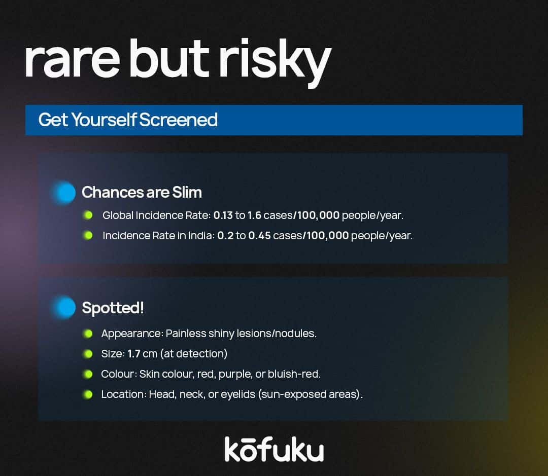
One tiny lump. One tiny nondescript lump, which could be anywhere - your face, hands, torso or in your fate - one tiny dime-sized lump is enough to make you say your prayers. This little bluish-red lump is less than two centimetres across, i.e. you won’t even know such a lump existed on your skin until you see it, and it visibly freaked you out.
But if you are on high alert, let me tell you that it’s justified. These lumps hurt a little less than your ex. And by that, I mean they don’t hurt at all. So why should you be bothered about them in the first place? Is it because all lumps are to be examined, or is it because you’re just paranoid?
Well, your paranoia is justified because this tiny lump on your skin could mean Merkel Cell Carcinoma. And, make no mistake - just because it’s a single lump doesn’t mean the cancer won’t spread. It will spread like gossip to your lymph nodes, liver, lungs and bones.
Merkel Cell Carcinoma - a rare but aggressive variety of skin cancer that can be life-threatening. This cancer impacts the outer layer of the epidermis or the skin. It often spreads to other lymph nodes and organs. What are the risk factors? Exposure to UV light, poor immunity, age and viral infection. And terrible luck. Just like cervical cancer.
What are Merkel cells?
Merkel cells are situated in your epidermis, which is the top layer of the skin. These are neuroendocrine cells that function in the nervous and endocrine systems. Sitting near nerve endings that provide a sense of touch, these cells were first described by Friedrich Sigmund Merkel in the late 1800s.
A Little History Before Anything Else -
Friedrich Sigmund Merkel was a German histopathologist who first described the Merkel cell in 1875. Fixing and staining the skin of geese and ducks, he demonstrated touch cells in pigs' snouts. These cells were at the dermo-epidermal junction, near myelinated nerve fibres. Merkel concluded that such cells acted as mechanoreceptors in all animals.
Cyril Toker later contributed to the study of this area in 1972 and had a research paper called “Trabecular Carcinoma of the Skin”. Later, immunohistochemistry and electron microscopy studies highlighted that such tumours emerge from the Merkel cell.
Causes
If you are unlucky enough to be exposed wrongly to the sun, and your immune system takes a few days off, then you are at risk of Merkel cell carcinoma. But don’t get your hair in a bunch. Having a risk factor doesn’t mean that you will get cancer; not having a risk factor doesn’t mean that you will be spared. What is the best way to find out? Speak to a doctor.
In any case, risk factors for Merkel Skin Carcinoma are -
-
Exposure to a lot of natural sunlight.
-
Exposure to artificial light, like that which comes from tanning beds, or psoralen and ultraviolet A (PUVA) therapy for psoriasis.
-
Having your immune system compromised by disease, like chronic lymphocytic leukaemia or Human Immunodeficiency Virus infection.
-
Ingesting medicines that make your immune system less active, like after having an organ transplant.
-
Having a history of other types of cancer.
-
Being older than 50 years of age.
Signs of Merkel Cell Carcinoma
This cancer originates, like all other cancers, from a tumour. Tumours from Merkel Cell Carcinoma usually crop up on sun-exposed areas of the skin. You might find a shiny or pearly lump on a particular area of skin that is often exposed to the sun. These infernal lumps might crop up anywhere.
Your face, neck, arms or eyelids. People who have darker skin might get these tumours on their legs. This lump might suddenly appear on your torso like Tinder notifications for people on the favourable side of the age spectrum. And you know what’s worse? This lump might also break open into a sore or a wound.
This lump is
-
About the dimensions of a dime and how it is growing faster than civilisation.
-
Dome-shaped or raised.
-
Itchy
-
Firm
-
Just like a pimple or an insect bite
-
The colour of your skin, or red, bluish-red or purple. This sly lump might be pink, red or purple, but it might also blend with the surrounding skin, making it extremely difficult to spot.
-
Sometimes, the tumour might become an ulcer or bleed as it grows.
-
The lump might be tender or sore.
-
As the disease worsens, it might spread to nearby lymph nodes, resulting in swelling - the first sign of trouble.

Tests For Finding Out Merkel Cell Carcinoma
Once Merkel Cell Carcinoma has been diagnosed, a few tests are carried out to check whether the cancer has spread to other body parts. There is a process to determine whether the cancer has spread to other body parts - staging.
Staging is essential to plan treatment. Just like treatment for ovarian cancer requires planning, so does the treatment for Merkel Cell Carcinoma.
CT Scan (CAT SCAN) This procedure makes several detailed pictures of areas inside our body from different angles. A computer linked to an X-ray machine takes these photos. A dye might be injected into a vein or swallowed to enable the organs or tissues to show up clearly.
A CT scan of the thoracic cavity might be used to find small-cell lung cancer or Merkel Cell Carcinoma that has metastasised.
A CT scan of the head and neck might be done to find Merkel Cell Carcinoma that has spread to the lymph nodes. This is called computed tomography, computerised tomography or computerised axial tomography.
PET (Positron Emission Tomography Scan)
This procedure finds malignant tumours in the body. A tiny amount of radioactive glucose is injected into a vein. The PET scanner goes around the body and takes pics of where the glucose is being used. Malignant cells show up brighter in the photo because of more activity, and they take up more glucose than regular cells.
Lymph Node Biopsy
There are many kinds of lymph node biopsy implemented to stage Merkel Cell Carcinoma -
- Sentinel lymph node biopsy -
Removing sentinel lymph nodes during surgery - this lymph node is the first in a group of lymph nodes to get lymphatic drainage from the primary tumour. This is the first lymph node the cancer might spread to from the primary tumour. It is injected with a radioactive substance or a blue dye, which flows through the lymph ducts to the lymph nodes.
The first lymph node that receives the substance or dye gets removed. A pathologist sees this tissue under a microscope to check for cancer cells. If they aren’t found, it isn’t required to remove more lymph nodes. A sentinel lymph node is found in more than one group of nodes.
- Lymph node dissection -
This is a surgical procedure in which the lymph nodes get removed, and a tissue sample is checked under a microscope to detect cancer. There’s regional lymph node dissection, where some lymph nodes in the tumour area get removed.
There’s radical lymph node dissection, where most or all the lymph nodes in the tumour area get removed. This is called a lymphadenectomy.
- Core needle biopsy -
This procedure removes tissue samples using a wide needle. A pathologist then scans this under a microscope to check for cancer cells. There are two types: core needle biopsy and fine needle aspiration biopsy.
- Fine-needle aspiration biopsy -
A thin needle is used to remove tissue samples, which are then viewed under a microscope to check for cancer cells.
Immunohistochemistry
This lab test uses antibodies for particular antigens in a tissue sample. The antibodies are mostly linked to an enzyme or a fluorescent dye. After these antibodies bind to a specific antigen in the tissue sample, the enzyme or dye gets activated, and the antigen can be viewed under a microscope. This kind of test finds cancer and tells one type from another.

Treatment Options for Patients with Merkel Cell Carcinoma
There are various kinds of treatments for patients afflicted with Merkel Cell Carcinoma. Some are standard, while others are tested in clinical trials. Treatment clinical trials are research studies that are supposed to improve present treatments or get new information on new treatments for cancer patients.
If clinical trials show that a new treatment has trumped standard treatment, the new treatment might become the standard treatment. Patients may want to think about participating in a clinical trial. They are open only to patients who haven’t commenced treatment.
There are four kinds of standard treatment -
Surgery
There are one or more surgical procedures to treat Merkel Cell Carcinoma.
- Wide Local Excision -
The cancer gets cut from the skin and some of the surrounding tissue. A sentinel lymph node biopsy might occur during the exhaustive local excision procedure. If cancer is present in the lymph nodes, a lymph node dissection might be carried out.
- Lymph node dissection -
In this surgical procedure, lymph nodes are removed, and a tissue sample is checked for signs of cancer. There are regional lymph node dissections in which some lymph nodes are removed or a radical lymph node dissection where most or all lymph nodes near the tumour get removed. This procedure is called a lymphadenectomy.
Radiation Therapy
This treatment implements high-energy X-rays or other radiation methods to kill cancer cells and prevent growth. External radiation therapy depends on a machine outside the body to send radiation to the area afflicted with cancer. It is used for treating Merkel Cell Carcinoma and palliative treatment.
Chemotherapy
This cancer treatment depends on drugs to stop cancer cell growth. It kills these cells and prevents division. Taken orally or injected into a vein or muscle, the medicines enter the bloodstream and reach cancer cells everywhere.
Immunotherapy
This treatment uses the patient’s immune system to battle cancer. Substances made by the body or in a laboratory are used to restore, direct or boost the body’s natural cancer defences. This is a kind of biological therapy.
Certain immune cells, like T cells and specific cancer cells, have particular proteins, named checkpoint proteins, on their surface that ensure immune responses are checked. When cancer cells have many proteins, T cells cannot affect them. Immune checkpoint inhibitors block such proteins, and T cells can kill cancer cells quickly.
There are two types of immune checkpoint inhibitor therapy -
- PD-1 and PD-L1 inhibitor therapy -
PD-1 is a protein found on T cell surfaces that checks the body’s immune responses. It can also be found in certain kinds of cancer cells. If PD-1 attaches to PD-L1, it prevents the T cell from killing the cancer cell. PD-1 and PD-L1 inhibitors prevent PD-1 and PD-L1 proteins from attaching.
This permits T cells to kill cancer cells. Pembrolizumab is a kind of PD-1 inhibitor, and avelumab is a kind of PD-L1 inhibitor used in treating advanced Merkel Cell Carcinoma. Then comes Nivolumab, a PD-1 inhibitor used to treat advanced Merkel Cell Carcinoma.
- CTLA-4 inhibitor therapy -
CTLA-4 is a protein on T cell surfaces that checks our immune responses. If CTLA-4 attaches to another protein called B7 on a cancer cell, it prevents the T cell from killing the cancer cell. CTLA-4 inhibitors attach to CTLA-4 and allow T cells to kill cancer cells. Ipilimumab is a kind of CTLA-4 inhibitor that is being researched for treating advanced Merkel Cell Carcinoma.
In conclusion, no matter how you choose treatment, Merkel Cell Carcinoma cannot be ignored. This cancer is sporadic and impacts very few people worldwide. It can be treated, however, using several different treatment options. If you notice a tiny lump anywhere, don’t panic. Get a biopsy done just to be sure, and if cancer surfaces, get treatment to remain cancer-free.
FAQs
I have a lump on my hand. Should I get a biopsy for Merkel Cell Carcinoma?
If the lump is bluish-red, you should get checked for Merkel Cell Carcinoma.
What is Merkel Cell Carcinoma?
Merkel Cell Carcinoma is a rare, aggressive form of skin cancer that develops from Merkel cells, specialised cells found in the skin responsible for the sensation of touch.
How common is Merkel Cell Carcinoma?
Merkel Cell Carcinoma is considered very rare. It accounts for less than 1% of all skin cancers.
What are the symptoms of Merkel Cell Carcinoma?
The primary symptom of MCC is the appearance of a painless, firm, and rapidly growing nodule or bump on the skin, often with a reddish or bluish colour.
Is Merkel Cell Carcinoma curable?
If detected early and treated appropriately, Merkel Cell Carcinoma can be curable. However, it grows and spreads quickly, so the prognosis depends. If diagnosed early, the chances of being cured are better.


Anatomy of Cancer: Understand Its Development

The History, Impact, and Importance of Vaccines Worldwide

Hodgkin’s vs Non-Hodgkin’s Lymphoma: Key Differences


