Ultrasound of Whole Abdomen: Test, Price, Procedure & Report Guide

Introduction
Ever been advised by your doctor to go and get an ultrasound of your whole abdomen, and think to yourself - how and why can this single test provide so much information? There are many reasons why linear transducer sonography has become one of the most popular diagnostic investigations in India. This includes its usefulness, low risk, speed, and relatively lower cost compared to CT or MRI scans.
Patients are usually advised to take this test to find out problems with the digestive tract, kidneys, liver, or reproductive organs. Understanding the details, such as how the procedure is performed, its price, how to read an ultrasound report of the whole abdomen, and what diseases can be detected by ultrasound, can transform the process and make it less stressful and more valuable.
What is an Ultrasound of the Whole Abdomen?
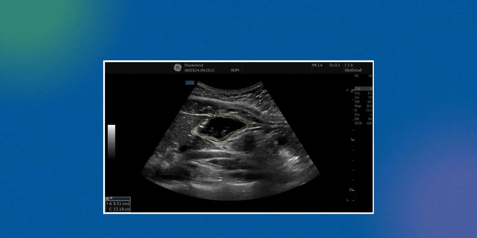
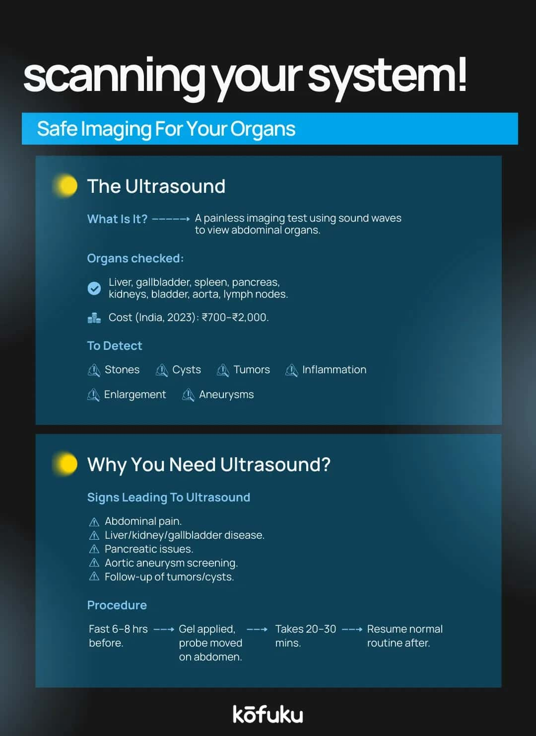
A non-invasive imaging test, such as an ultrasound of the whole abdomen, or whole abdomen USG, uses sound waves to form pictures of the organs of the abdomen. The technique does not use radiation, as in the case of X-rays, and is therefore safe for both adults and children.
This test checks some of the significant abdominal structures such as the liver, the gallbladder, pancreas, spleen, kidneys, bladder, intestines, and, occasionally, even the reproductive organs. Whole abdomen scanning is typically advised in cases where patients come with long-lasting abdominal pains, abdominal swelling, sudden and unwarranted weight loss, and possible internal infections.
Types of Abdominal Ultrasound Tests
Not all patients need a complete scan. Depending on the symptoms, doctors may indicate or request certain forms of sonography, such as abdominal and pelvic tests. A few of the common variations are:
USG Upper Abdomen Explained
A USG upper abdomen or an ultrasound of the upper abdomen is performed to evaluate the liver, gallbladder, bile ducts, spleen, pancreas, and kidneys. It is generally ordered in situations where the patient complains of jaundice, gallstones, or a suspected fatty liver.
Tumours, abscesses, and infections occurring in the organs of the upper abdomen can also be detected in this test.
USG Test for Pelvis
Pelvic sonography is used to check the reproductive organs, which include the uterus, ovaries, fallopian tubes, among women, and the prostate gland among men. It also evaluates the urinary bladder and peri-bladder tissue. An abdominal and pelvic sonography may be suggested for patients complaining of irregular menstrual cycles, fertility problems, or pelvic pain.

How the Ultrasound Procedure is Done
The process is easy and generally takes 15-30 minutes. A trained technician or radiologist applies a gel to the abdomen to enhance the transmission of sound waves. A transducer, a handheld device, is then placed and moved on the surface of the abdomen.
The reflected lines of sound create real-time images on a monitor, which are then recorded as the ultrasound report of the whole abdomen. The procedure is painless, but patients may experience mild pressure with the movement of the probe on tender regions.
Colour Doppler in Abdominal Ultrasound
There are cases when physicians need to have detailed data concerning the blood flow in abdominal vessels. Here, Colour Doppler Ultrasound comes into the picture. It outlines the colour of arteries and veins to indicate the state of blood flow through the intestinal organs.
A Colour Doppler test is particularly useful for detecting blockages in abdominal blood vessels, evaluating liver diseases, or monitoring patients with kidney transplants.
Colour Doppler Test Price
The Colour Doppler Test cost in India falls in the range of ₹1200-₹3500 per test, depending on the city and hospital, as well as whether the test is partial or for the whole abdomen.
Abdominal Ultrasound Price
Abdominal ultrasound can be used broadly because it is cheap. The cost of an abdominal ultrasound in India would lie between ₹500 and ₹2000 in the majority of diagnostic centres.
A privately operated hospital might prove to be more expensive, but government facilities can offer subsidised fees. Given this, ultrasound remains one of the most cost-effective imaging tools for long-term monitoring.

What Diseases Can Be Detected by Ultrasound?
What diseases can be detected by ultrasound? Some of the most common findings include:
- Liver problems: Fatty liver, hepatitis, cirrhosis, liver abscess, and tumours.
- Gallbladder disorders: Gallstones, infections, and inflammation.
- Pancreatic diseases: Pancreatitis, cysts, or masses.
- Kidney issues: Stones, infections, cysts, and hydronephrosis.
- Bladder conditions: Tumours, infections, and stones.
- Reproductive organs: Ovarian cysts, fibroids, and prostate enlargement.
- Abdominal infections: Abscesses and fluid accumulation (ascites).
How to Read an Ultrasound Report of the Whole Abdomen
Interpretation of an ultrasound report of the whole abdomen requires medical knowledge; however, patients can identify key terms. Reports tend to describe organ size, the structure of the organs, the existence of cysts, stones, or abnormal growths.
If the report mentions terms like “hyperechoic” or “hypoechoic,” these describe how tissues reflect sound waves. Doctors interpret these findings to confirm or rule out disease.
USG of Whole Abdomen Report Format
A typical USG of the whole abdomen report includes:
- Patient details and scan date
- Organ-wise findings (liver, gallbladder, pancreas, spleen, kidneys, bladder, intestines)
- Any abnormalities detected (stones, masses, fluid, inflammation)
- Impression or conclusion by the radiologist
Whole Abdomen Scanning – When and Why it’s Recommended
Whole abdomen scanning is recommended when the symptoms are ambiguous or apply to more than one area of the abdomen. It is particularly helpful in situations of:
- Persistent abdominal pain
- Unexplained fever
- Digestive problems not explained by blood tests
- Follow-up of chronic liver or kidney diseases
- Suspected abdominal tumours
Risks and Safety of Abdominal Ultrasound
Ultrasound can be regarded as the safest imaging technique. It neither utilises radiation nor is it harmful to pregnant women. There are no long-term dangers, as is the case with CT scans.
Preparing for an Abdominal Ultrasound
Proper preparation ensures accurate imaging. Patients are often advised:
- Fasting: 6-8 hours before scan for liver, gallbladder, or pancreas.
- Hydration: Drink water before a bladder ultrasound.
- Avoid gas-forming foods: This prevents bloating that may obscure images.
- Follow the specific doctor’s advice, especially for diabetic or pregnant patients.
Conclusion
An ultrasound of the whole abdomen is a cost-effective, safe and valuable tool in the detection of numerous abdominal diseases. This quick and non-invasive test not only provides fast and accurate information, which is ultimately used to inform treatments, but it has also been used to diagnose liver problems, kidney stones, and other conditions. Be it USG upper abdomen or sonography abdomen and pelvis or whole body scan with colour doppler, each is a vital test.
To make appropriate health decisions, patients must also know the price of an abdominal ultrasound and the expected contents of their USG of the whole abdomen report. Visiting an abdominal expert is also relevant to her problems to get an accurate diagnosis during the consultation.

FAQs
Q. What is the difference between the USG whole abdomen and the upper abdomen?
A. A USG whole abdomen covers all abdominal and pelvic organs, while an upper abdomen ultrasound focuses mainly on the liver, gallbladder, pancreas, spleen, and kidneys.
Q. How much does an abdominal ultrasound cost in India?
A. The cost of an abdominal ultrasound in India usually ranges between ₹500 and ₹2,000, depending on the city, diagnostic centre, use of Colour Doppler, and additional specialist interpretation.
Q. How to read an abdominal ultrasound report?
A. An abdominal ultrasound report includes organ sizes, shapes, and abnormalities like stones or cysts. Patients should review impressions but rely on their doctor for accurate interpretation and clinical correlation.
Q. What diseases can be detected by an ultrasound of the whole abdomen?
A. Ultrasound of the whole abdomen detects liver disease, gallstones, kidney stones, pancreatic inflammation, ovarian cysts, bladder tumours, abdominal infections, fluid accumulation, and sometimes cancers. However, small or early tumours may need CT/MRI.
Q. Is Colour Doppler included in abdominal ultrasound?
A. Not always. A standard abdominal ultrasound may exclude Colour Doppler, which is typically recommended separately for evaluating blood flow in abdominal vessels. Pricing varies when Colour Doppler is added.
Q. How long does a whole abdomen ultrasound take?
A. A whole abdomen ultrasound usually takes 20 to 30 minutes. Additional time may be needed if Colour Doppler is used, or if complex conditions require detailed scanning.
Q. How to prepare for an abdominal and pelvic ultrasound?
A. Preparation includes fasting for 6-8 hours for abdominal scans and drinking water to keep the bladder full for pelvic scans. Follow the doctor’s instructions carefully for accuracy.
Q. Can an abdominal ultrasound detect cancer?
A. Yes, an abdominal ultrasound can detect some cancers, such as liver or kidney tumours. However, small or early-stage cancers often require further confirmation with CT, MRI, or biopsy.

A Complete Guide to Women’s Health: From Body Metrics to Health Issues in India

Beyond the Bump: Why Diabetes in Pregnant Women Demands Your Attention Now

Impact of Alcohol on Women’s Health Is More Than Men


