Home
Blogs
Wellness Corner
Understanding Liver Hemangioma: Definition, Causes & Importance of Early Diagnosis
Understanding Liver Hemangioma: Definition, Causes & Importance of Early Diagnosis

Introduction
Have you ever made a regular visit to the doctor only to find something unexpected has occurred in the ultrasound report, maybe a strange spot on your liver? This is generally diagnosed as a liver hemangioma. It is common in India and is generally found in many individuals who have abdominal pain, indigestion, or other unrelated problems during imaging.
As the diagnosis is made, it usually instils fear in the patient as it is automatically subsequent with cancer or severe liver disease. Liver hemangioma is mostly a benign condition, and proper knowledge of the same is pivotal in avoiding undue anxieties and in proper medical care.
This article explores what a liver hemangioma is, its causes, radiology findings, dietary advice, and the importance of accurate diagnosis and timely intervention.
What Is a Liver Hemangioma?
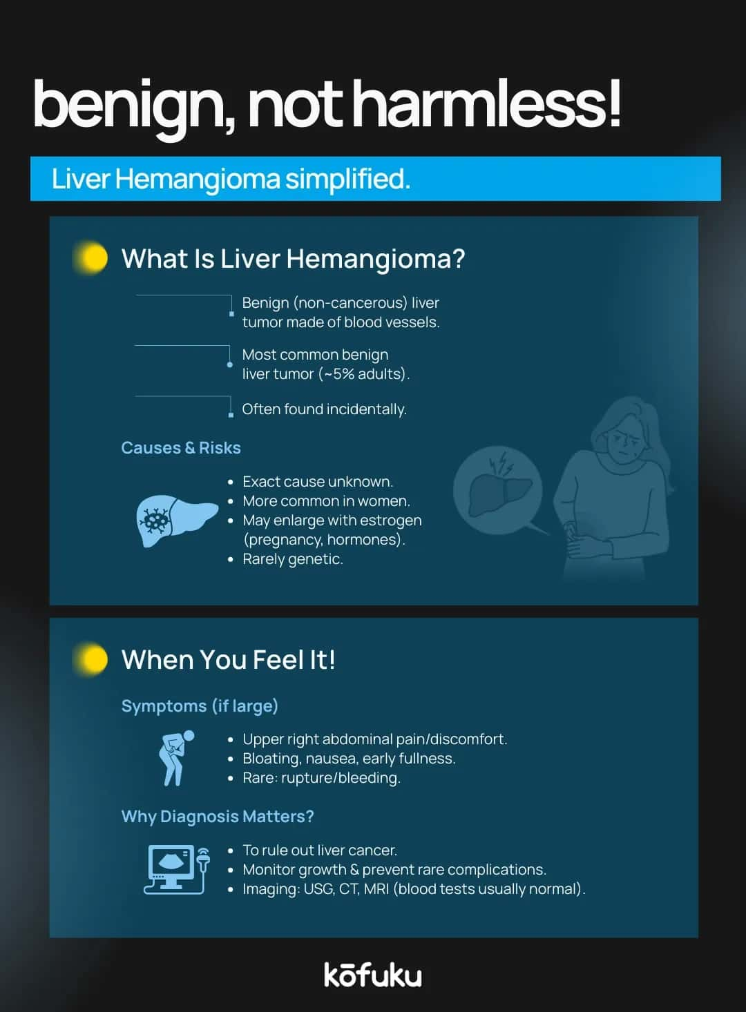
A liver hemangioma is a benign tumour of the liver consisting of a cluster of deformed blood vessels. It can be mistaken for other liver lesions, such as liver SOL (space-occupying lesion) or liver hematoma, but it still has its peculiarities. Oftentimes, liver hemangiomas are asymptomatic and small in size, and they are found randomly when imaging tests are performed.
In contrast to cancerous tumours, hemangiomas will infrequently grow aggressively or spread. A large number of patients spend the whole time without the requirement of any treatment. But bigger hemangiomas can occasionally result in abdominal pain, nausea, or discomfort when pressing against other organs.
Liver Hemangioma Radiology: Ultrasound, CT, MRI Findings
Radiology plays a vital role in detecting and differentiating liver lesions. Since liver hemangiomas can mimic other conditions, imaging accuracy is essential.
Liver Hemangioma Ultrasound: What to Expect and Interpret
The first line of diagnostic investigations is an ultrasound. Hemangiomas in general appear on ultrasound as hyperechoic (bright) lesions, which can be confused with echogenic changes in the liver. Where lesions are small, their classical appearance suffices to make the diagnosis. CT and MRI offer more detail with larger or unusual lesions.
Hemangiomas have a nodular peripheral enhancement pattern that fills in centrally on contrast-enhanced CT or MRI. That is one of the significant distinguishing factors between malignant lesions.

Liver Hemangioma ICD-10 Codes: Accurate Coding for Diagnosis and Billing
The ICD-10 code D18.0 identifies benign liver hemangiomas, ensuring precise medical documentation, accurate billing, and standardised diagnosis for effective patient management and healthcare reporting.
Differential Diagnosis: Liver Sol, Hematoma, and Other Liver Lesions
A liver hemangioma must be differentiated from other space-occupying lesions (SOL).
-
Liver SOL: A broad term covering any mass or growth in the liver, which could be benign or malignant.
-
Liver Hematoma: Refers to a collection of blood in the liver, usually due to trauma or injury. Unlike hemangiomas, hematomas often resolve over time.
-
Correlating with Radiology: A detailed hemangioma liver radiology assessment helps rule out malignancy, cysts, or abscesses.
Liver Hemangioma Diet: Foods and Lifestyle for Liver Health
Although there is no specific liver hemangioma diet, starting with liver-friendly nutrition is advisable. Physicians tend to advise:
- A balanced diet rich in vegetables, fruits, and whole grains.
- Avoiding excess alcohol, which can strain the liver.
- Limiting high-fat and fried foods, which may worsen liver stress.
- Staying hydrated and including antioxidant-rich foods.
Liver Function Tests Abnormal ICD-10 and Interpretation in Liver Hemangioma
Liver hemangiomas do not generally lead to changes in liver functioning. But in that case, when an unusual level of liver enzymes is detected in blood tests, this can indicate concomitant liver problems: fatty liver, hepatitis, or cirrhosis.
- Liver enzymes abnormal ICD-10: R74.8 (abnormal levels of other specified enzymes).
- In the presence of a hemangioma, abnormal liver function tests (LFTs) usually require further investigation to rule out additional diseases.

When to Consider Treatment: Indications and Available Options for Liver Hemangioma
Most liver hemangiomas do not require treatment. However, medical intervention becomes necessary when:
- The lesion is larger than five cm and causes pain or pressure.
- There are complications such as bleeding or rupture (rare).
- Diagnostic uncertainty exists despite advanced imaging.
Interventional Radiology and Intravenous Treatments for Liver Hemangioma
There are specific circumstances in which interventional radiology methods, like arterial embolisation, are implemented to reduce the size of a hemangioma. It seldom needs surgery, which can be contemplated in very large or symptomatic instances. Innovative treatments, such as intravenous modes, are being studied but are not yet standard.
Nursing Care and Monitoring for Patients with Liver Hemangioma
Nurses are vital in the care provided to patients with liver hemangioma, especially when they need some procedures or follow-ups. Nursing care incorporates:
- Monitoring for abdominal pain, swelling, or signs of rupture.
- Supporting patients undergoing radiology tests and explaining procedures like ultrasound or MRI.
- Educating patients about diet, lifestyle, and regular follow-up.
- Keeping accurate documentation with ICD-10 coding for billing and record-keeping.

FAQs
Q. What is a liver hemangioma, and how is it diagnosed?
A. A liver hemangioma is a benign vascular liver tumour. Diagnosis typically involves imaging tests like ultrasound, CT scan, or MRI to confirm size, location, and vascular characteristics.
Q. What are the ICD-10 codes for liver hemangioma?
A. The ICD-10 code for liver hemangioma is D18.0. It classifies benign vascular neoplasms of the liver, aiding in medical documentation, billing, and standardised diagnosis coding.
Q. How does a liver hemangioma appear on ultrasound or MRI?
A. On ultrasound, it appears as a well-defined, hyperechoic lesion. MRI shows high signal intensity on T2-weighted images, highlighting its vascular structure, differentiating it from malignant liver lesions.
Q. What is the difference between a liver hematoma and a liver hemangioma?
A. A liver hematoma is a blood collection due to trauma, while a hemangioma is a benign vascular tumour. Hematomas usually resolve over time; hemangiomas are often stable and asymptomatic.
Q. What is the meaning of echogenic liver in ultrasound reports?
A. “Echogenic liver” indicates increased liver brightness on ultrasound, suggesting fatty infiltration, fibrosis, or other liver conditions. It describes tissue density, not necessarily the presence of tumours.
Q. What does “liver SOL” mean in radiology, and how is it related to hemangioma?
A. “Liver SOL” refers to a space-occupying lesion in the liver. Hemangiomas are a type of SOL, representing benign vascular masses detected through imaging studies.
Q. What diet is recommended for someone with a liver hemangioma?
A. A balanced diet rich in fruits, vegetables, lean proteins, and whole grains supports liver health. Avoid excessive alcohol, fatty foods, and hepatotoxic substances to reduce liver stress.
Q. Can liver hemangiomas cause abnormal liver function test results?
A. Most liver hemangiomas are asymptomatic and do not affect liver function tests. Rarely, very large hemangiomas may mildly alter enzyme levels or cause discomfort.

Fatty Liver in Women: Causes, Symptoms, and Stage-by-Stage Progression

Hepatoblastoma-Liver Cancer in Children

How Coffee Affects Your Liver and Kidneys: Benefits, Risks & Daily Use

Liver Transplant Donors: What You Need to Know

Liver Cancer Survival Guide: What You Should Know


